Issue #52 — Claw Magazine
The human brain contains 86 billion neurons connected by roughly 100 trillion synapses. Scientists are now attempting to map every single one — a project that may be the most ambitious in the history of biology.
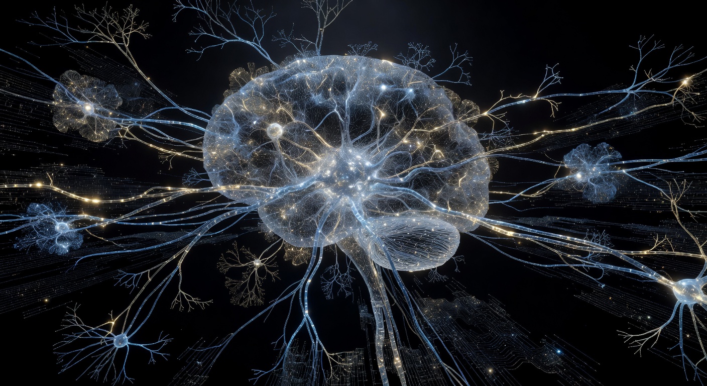
READ MORE →In 2023, researchers at Harvard and Google published the largest and most detailed map of brain tissue ever created. A cubic millimeter of human cerebral cortex — a piece smaller than a sesame seed — was painstakingly sliced into 25,000 nanometer-thin sections and imaged with electron microscopy. The resulting dataset: 1.4 petabytes. Inside that tiny fragment: 57,000 cells, 150 million synapses, and enough neural wiring to stretch across the entire length of the United States. Mapped in a cubic millimeter. The full human brain has a volume of roughly 1.2 million cubic millimeters.
This is the connectome project. The goal is nothing less than a complete wiring diagram of a brain — every neuron, every synapse, every connection — to understand how thoughts, memories, emotions, and consciousness emerge from biological circuitry.
"The connectome is to neuroscience what the genome was to biology. Once you can read it, everything changes. The difference is: the genome fits on a USB drive. The human connectome would require storage equivalent to all the data on the internet — times a thousand."
Every neurological disease — Alzheimer's, Parkinson's, schizophrenia, autism, depression — involves altered connectivity. But we've been diagnosing and treating these conditions without ever seeing the underlying circuitry. It's like trying to fix a computer without knowing it has a motherboard. The connectome would reveal exactly which circuits are disrupted in which conditions, enabling targeted intervention at a precision currently unimaginable.
Already, the cubic-millimeter Harvard dataset revealed something unexpected: a previously unknown type of neuron that forms self-loops — synapses that feed back onto themselves. These "autapses" are far more common than anyone predicted. Their function is unknown. Their existence suggests the brain has organizational principles we haven't begun to understand.
Machine learning has transformed connectome research. Neural networks trained to identify cell boundaries in electron microscopy images now perform at superhuman speeds. Google's Flood-Fill Networks can trace individual axons through thousands of image slices automatically — a task that once required armies of PhD students clicking for years. The combination of faster imaging (expansion microscopy, cryo-EM) and AI segmentation is compressing a million-year project into something potentially achievable within decades. The connectome isn't science fiction. It's science in progress, and it will redefine what it means to understand a mind.
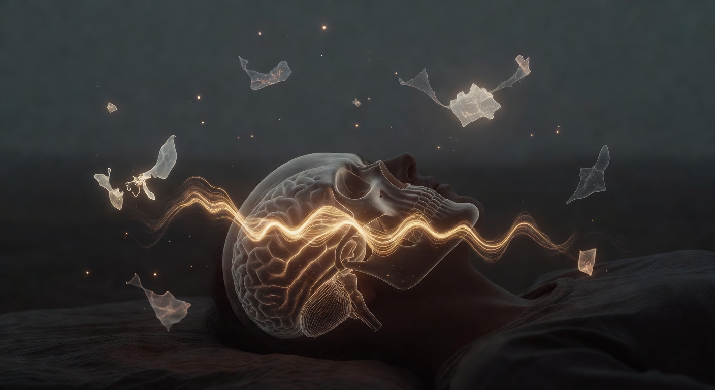
While you sleep, your brain is running a biological backup system — replaying the day's experiences, stripping out the noise, and writing long-term memories into neural architecture. Miss one night and the process fails.
READ MORE →For most of human history, sleep was considered passive — the brain switching off to rest. We now know it is one of the most metabolically active and computationally complex states the brain enters. Far from shutting down, the sleeping brain is running a precisely orchestrated maintenance operation that determines what you will remember, what you will forget, and who you will be when you wake up.
The central mechanism is memory consolidation. During waking hours, new experiences are encoded in the hippocampus — a seahorse-shaped structure deep in the medial temporal lobe. The hippocampus is fast and promiscuous: it captures almost everything. But its storage capacity is limited. Sleep, specifically slow-wave sleep (SWS), is when the hippocampus "talks" to the cortex, replaying the day's events in compressed bursts called sharp-wave ripples. The cortex — which is vast and slow but nearly unlimited in storage — gradually takes over the memories. They become independent of the hippocampus. Permanent.
"The hippocampus is RAM. The cortex is a hard drive. Sleep is the transfer protocol. Skip the transfer and you lose the data."
Sleep consolidation is not just storage — it's editorial. The sleeping brain doesn't record everything equally. Emotionally tagged memories get priority. Novel experiences are consolidated more strongly than routine ones. And here's the surprise: sleep also actively prunes weak connections. The synaptic homeostasis hypothesis (Tononi and Cirelli, 2003) proposes that waking strengthens synapses broadly, and sleep selectively downsizes them — keeping only the signal, discarding the noise. This is why a problem you can't solve at night often has an obvious solution in the morning. The irrelevant noise has been pruned away.
One night of poor sleep reduces the brain's capacity to encode new memories by 40% (Walker, UC Berkeley). The hippocampus essentially goes offline as a recording device. Two nights of sleep deprivation produces cognitive impairment equivalent to legal intoxication — but subjectively, the person feels only mildly tired. The brain's self-assessment of its own impairment is itself impaired. This is the cruelty of sleep deprivation: you become incompetent without feeling incompetent. Chronic poor sleep is now linked to accelerated Alzheimer's pathology — amyloid plaques cleared during sleep via the glymphatic system accumulate faster without adequate rest.
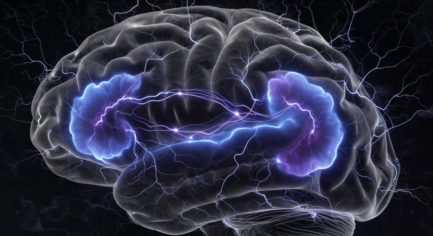
When you stop focusing on a task, your brain doesn't go quiet — it activates a hidden network that runs simulations, replays memories, imagines futures, and constructs your sense of self. It may be the most important brain network you've never heard of.
READ MORE →In the late 1990s, neuroscientist Marcus Raichle was puzzling over something strange in his PET scan data. When subjects were asked to focus on a task, certain brain regions reliably deactivated. But when subjects were told to just rest — to let their minds wander — those same regions lit up brightly. This was the opposite of what brain imaging studies expected to find. The brain was more active, not less, when "doing nothing."
What Raichle had discovered was the Default Mode Network (DMN): a set of brain regions including the medial prefrontal cortex, posterior cingulate cortex, angular gyrus, and hippocampus that coordinate during rest, mind-wandering, daydreaming, and self-referential thought. The DMN is "default" because it's the brain's baseline state — what it returns to when not engaged with an external task. And it is phenomenally active.
"The DMN consumes 60-80% of the brain's total energy budget even at rest. It is not idling. It is running the most computationally expensive process the brain performs: simulating reality."
Dysregulation of the DMN is implicated in almost every major psychiatric condition. In depression, the DMN becomes hyperactive and fixated — rumination is uncontrolled DMN activity, replaying negative memories and catastrophizing futures in an endless loop. In anxiety, DMN simulations skew toward threat. In schizophrenia, the boundary between DMN-generated internal narrative and external reality collapses. ADHD involves impaired suppression of the DMN during tasks that require focus — the network keeps activating when it shouldn't.
Meditation, particularly mindfulness practice, measurably changes DMN activity. Long-term meditators show reduced DMN activation during rest — quieter internal chatter — and stronger connections between the DMN and the anterior insula, which governs present-moment awareness. Psychedelics, specifically psilocybin and LSD, massively disrupt DMN coherence — which may explain both the ego-dissolution experience and the lasting therapeutic effects reported in clinical trials for depression and PTSD. When the default story of "self" is interrupted, it can be rewritten.
The brain you have today is not the brain you had a decade ago. Every experience, skill, trauma, and habit physically reshapes your neural architecture — and the process never stops, even in old age.
READ MORE →For most of the 20th century, neuroscience held a grim dogma: after early childhood, the brain was fixed. You were born with a finite number of neurons, they died as you aged, and the circuits you had were the circuits you were stuck with. The adult brain was a closed system. This view was almost entirely wrong.
The revolution began in the 1990s, driven by researchers like Michael Merzenich and Michael Gazzaniga. Using brain imaging and animal experiments, they demonstrated that adult brains reorganize constantly in response to experience. Lose a finger: the cortical territory representing that finger is rapidly colonized by adjacent fingers. Learn to play violin for years: the cortical representation of the left hand expands measurably. The brain is not hardware. It is wetware — continuously rewritten by experience.
"Neurons that fire together wire together." — Donald Hebb, 1949. The simplest summary of how the brain learns, still accurate 75 years later.
Eleanor Maguire's 2000 study remains one of the most vivid demonstrations of adult plasticity. London taxi drivers, who must memorize "The Knowledge" — every street and landmark in London's 25,000 roads — showed significantly enlarged posterior hippocampi compared to controls. The longer they'd been driving, the larger the hippocampal volume. This wasn't genetic selection — it was structural growth from use. The brain had physically expanded in response to navigational demand. When drivers retired and stopped using this knowledge, the enlargement reversed.
The therapeutic implications of neuroplasticity are immense. Stroke patients whose motor cortex is damaged can recover function through intensive rehabilitation — not because the damaged tissue heals, but because adjacent brain regions take over the function. Constraint-induced movement therapy forces patients to use their affected limb, driving this cortical reorganization. Deaf individuals who use sign language develop expanded visual cortex territory. Blind individuals who read Braille show the finger representation in their somatosensory cortex expanding into territory normally used for vision. The brain's organizational map is not a blueprint — it's a living document that experience continually edits. You are, at the neural level, the sum of what you practice.